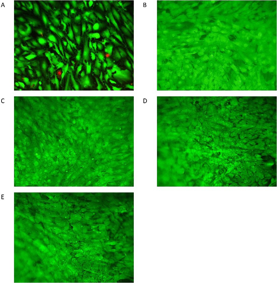
Fig. 2. Representative LIVE/DEAD staining, recorded with a Bio Zero 8100 fluorescence microscope from Keyence, 20x magnification, 1.5x10⁵ haSMC were grown on all displayed CellDrums. Due to the curvature of the CellDrum membrane, only a small part of the specimen is in the focal plane. (A) Reference LIVE/DEAD staining of the haSMC monolayer on the CellDrum. Functional testing of the staining, cell damage was induced by excessive bi-axial mechanical stretching. Intact cells are shown in green and damaged cells in red fluorescence. (B) Control, without the addition of active ingredient (C) Specimen after 30 minutes treatment with 1µM Bay K8644. (D) CellDrum after 30 minutes treatment with 1µM nifedipine (E) Cell monolayer after 30 minutes treatment with 1µM verapamil.